Winners of the 2023 Nikon Small World Microscopy Photography Contest
ArabiaWeather - The Nikon Small World 2023 Microscopic Photography Competition reflects the diversity of photographic subjects in all its shapes and sizes, as everything can be photographed, from vast natural scenes to small objects, and everything imaginable in between. However, the small microscopic world and its unique details often seem to Ignored, despite its visual splendor, photomicrography, or optical microscopy, is a specialized technique that links scientific photography with visual creativity.
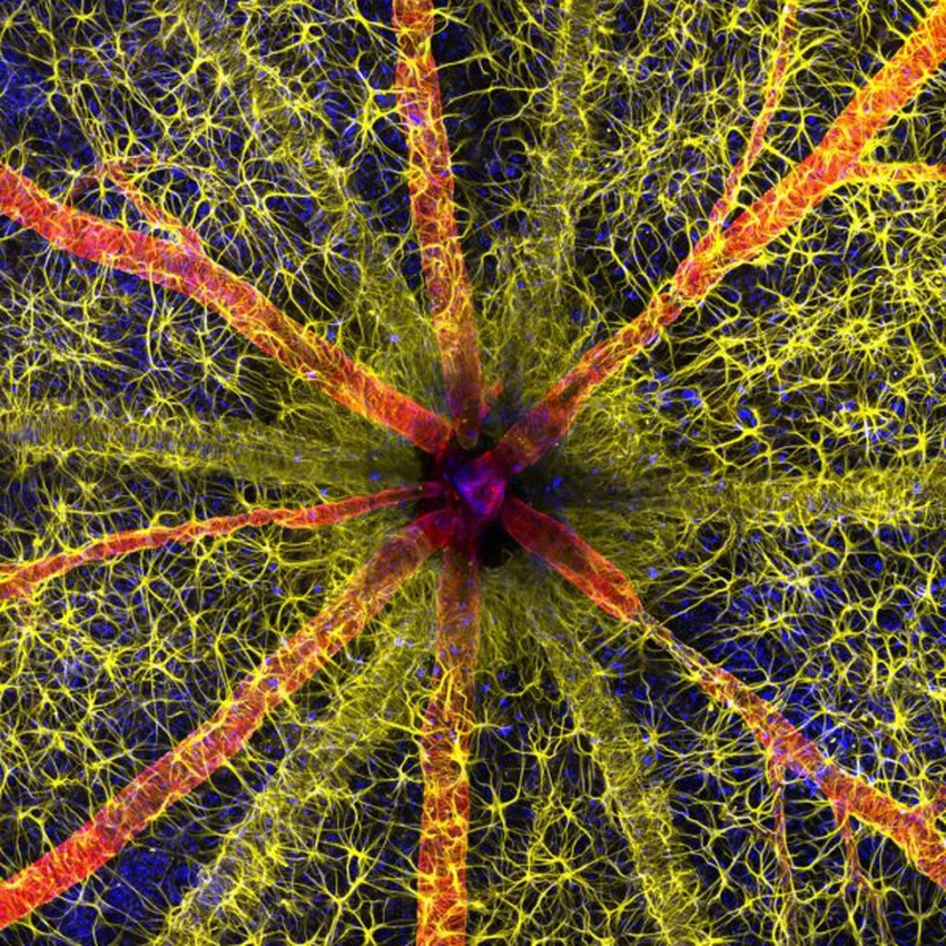
After forty-nine years of history, Nikon has announced the winners of its 2023 Small World Photo Contest and awards this year's grand prize to photographer Hassanein Gambari with the help of Jayden Dixon of the Lions Eye Institute for his stunning image of the optic nerve head of a rodent.
Here are the 10 best images from this year's competition, with information about the artist and details about the zooming and shooting techniques used. If you'd like to view the full gallery of this year's best images, you can visit the Nikon Small World photo gallery website.
first place
Rodent optic nerve head showing astrocytes (yellow), contractile proteins (red), and retinal blood vessels.
Artists: Hasnain Gambari, Jayden Dixon (Lyons Eye Institute, Department of Physiology and Pharmacology: Perth, Western Australia, Australia)
Magnification: 20X (objective lens magnification)
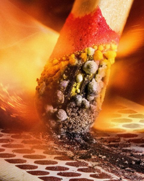
Second place
The ignition of the match due to friction on the surface of the box
Artists: Uli Bielefeldt (Macroving: Cologne, North Rhine-Westphalia, Germany)
Magnification: 2.5X (objective lens magnification)
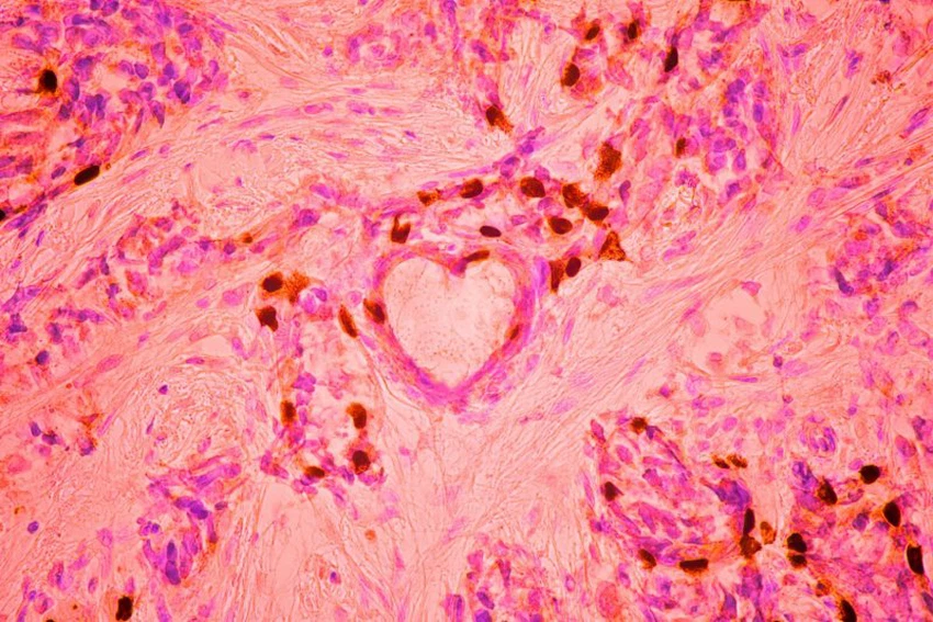
the third place
Breast cancer cells
Artists: Malgorzata Lisowska (Independent Value-Based Healthcare Consultant: Warsaw, Mazowiecki, Poland)
Magnification: 40X (objective lens magnification)
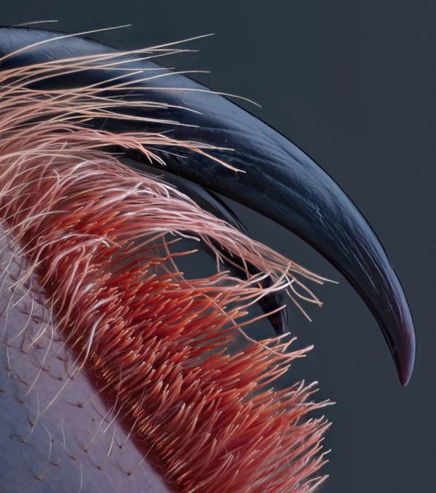
fourth place
Poisonous fangs of small tarantulas
Artists: John Oliver Dohm (Medienbunker Production: Bendorf, Rheinland Pfalz, Germany)
Magnification: 10X (objective lens magnification)
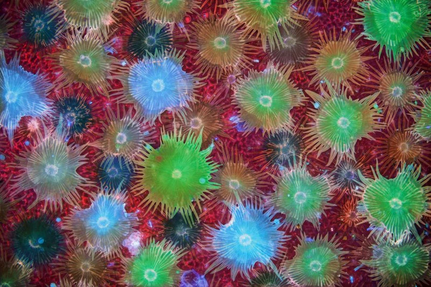
Fifth place
Autofluorescent defensive hairs cover the surface of Eleagnus angustifolia leaves exposed to ultraviolet radiation.
Artists: Dr. David Maitland (www.davidmaitland.com: Feltwell, Norfolk, United Kingdom)
Magnification: 10X (objective lens magnification)
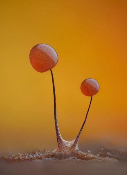
Sixth place
Slime mold (Comatricha nigra) that displays filamentous fibers through its transparent periphery
Artists: Timothy Boehmer (WildMacro: Vacaville, CA, USA)
Magnification: 10X (objective lens magnification)
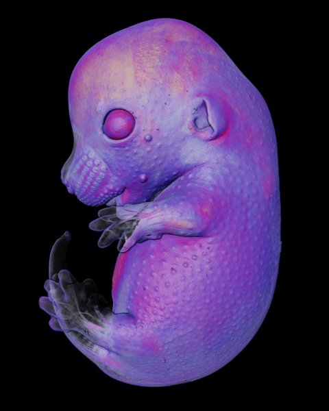
Seventh place
Mouse embryo
Artists: Dr. Gregory Temin, Dr. Michel Milinkovic (University of Geneva, Department of Genetics and Evolution: Geneva, Switzerland)
Magnification: 4X (objective lens magnification)
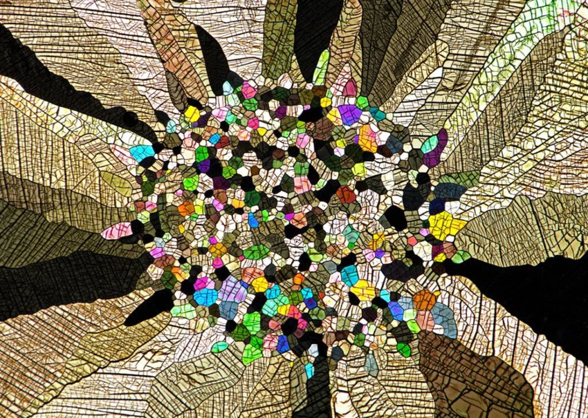
Eighth place
Caffeine crystals
Artists: Stefan Eberhard (University of Georgia: Athens, Georgia, USA)
Magnification: 25X (objective lens magnification)
Ninth place
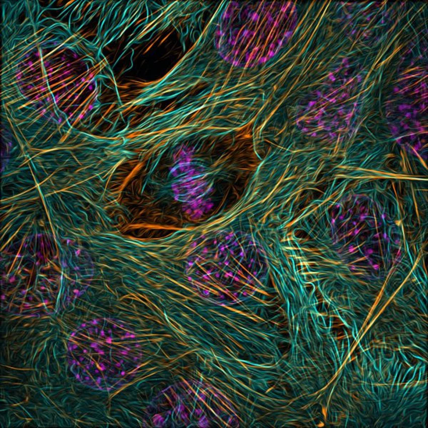
Cytoskeleton of dividing myoblast; Tubulin (cyan), F-actin (orange), nucleus (magenta)
Artists: Vaibhav Deshmukh (Baylor College of Medicine, Department of Molecular Physiology and Biophysics: Houston, TX, USA)
Magnification: 63X (objective lens magnification)
Tenth place
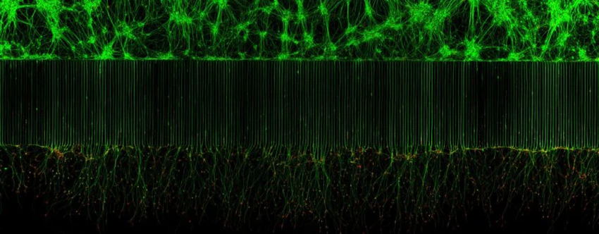
Motor neurons grown in a microfluidic device to separate cell bodies (top) and axons (bottom). Green - microtubules. Red - growth cones (actin)
Artists: Melinda Beccari, Dr. Don W. Cleveland (University of California, San Diego, Department of Cellular and Molecular Medicine: La Jolla, CA, USA)
Magnification: 20X (objective lens magnification)
Know also:
The sixth basic taste under the tongue... What is it?
Sources:
Arabia Weather App
Download the app to receive weather notifications and more..


